Bioinspired soft sensors and wearables for biomedical applications

Ajay Kottapalli: Associate Professor and Chair of Bioinspired MEMS and Biomedical Devices Group at the University of Groningen
This talk will delve into the fascinating realm of biomimicry, exploring how the mechanosensory designs of biological sensors observed in nature can be ingeniously transposed into artificial polymer sensors and flexible electronic devices. Ajay Kottapalli’s research develops flexible electronic devices, artificial skin sensors, electronic tattoos and wearable sensors for tracking motion, monitoring physiological parameters (sweat sensing), and enhancing human-machine interactions. Flexible nanomaterial-based sensors, encompassing piezoresistive, piezocapacitive, and piezoelectric functionalities, were fabricated and seamlessly integrated into the Internet of Things (IoT), showcasing compelling demonstrations of their biomedical sensing and monitoring applications. The talk will elucidate the process of translating these biological inspirations into practical applications, particularly focusing on their integration into biomedical sensing and healthcare devices.

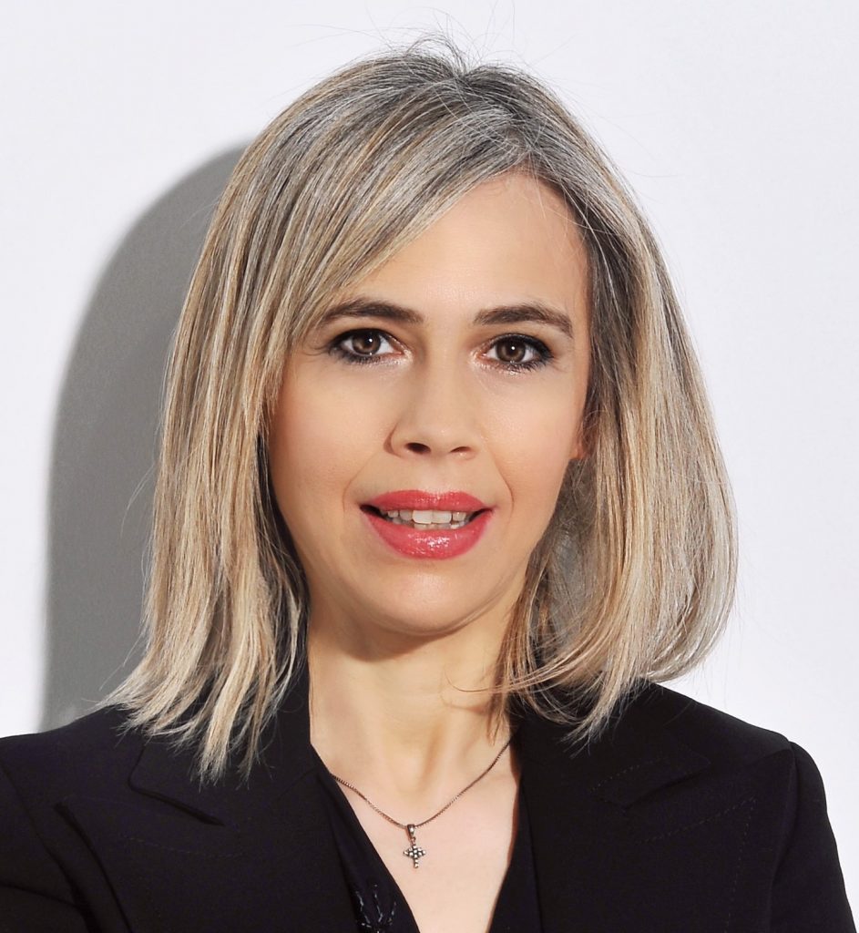
Distinguished lecturer of IEEE EMBS by Spyretta Golemati – Associate Professor of Biomedical Engineering in the Medical School of the National and Kapodistrian University of Athens, Greece
Using sound to hear, image, and treat cardiovascular function
Cardiovascular structures, including the heart and vessels, are sources of sound. Blood flow in arteries, as well as around heart valves, has unique acoustic features, which are altered in the presence of disease. Such features can be assessed with acoustic sensors (microphones and accelerometers), which, going beyond the stethoscope, the conventional method for assessing heart sounds, allow mapping of the acoustic phenomena of extended tissue areas. In addition to this passive approach of hearing cardiovascular sounds, ultrasound, i.e. sound with frequencies higher than the upper audible limit of human hearing, can be used to image cardiovascular tissues. Ultrasound images represent the result of the interaction (i.e. the echoes) of ultrasound beams directed to tissues, and are widely used in clinical diagnosis and image-guided therapy. Coupled with advanced image analysis methods, conventional ultrasound imaging can yield novel markers of cardiovascular function, including morphological, textural, and mechanical indices that describe complex physiological phenomena. The combination of such markers with machine learning can predict and justify adverse cardiovascular events, towards an in-depth understanding of vascular physiology and pathophysiology and a personalised patient stratification.